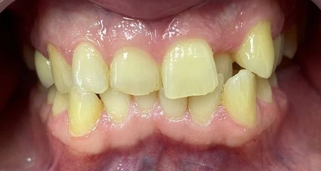
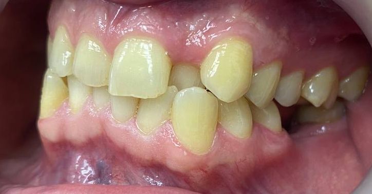
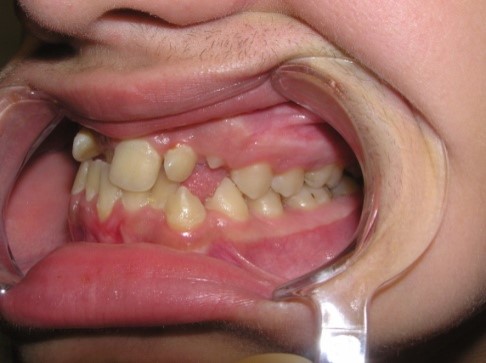
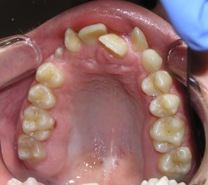
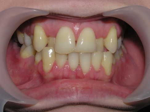
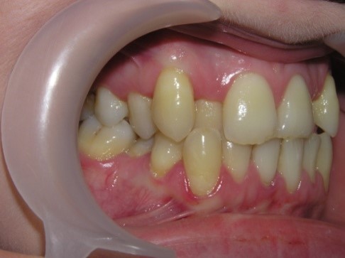
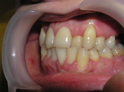
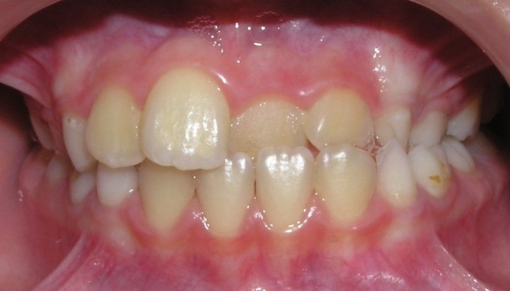
Palatal teeth position
Palatal position of teeth is
characterized by the eruption of one tooth or a group of teeth outside the
dental arc on the palatal side. So, most often, incisors or second premolars
eruption.
The frequency of palatal position of
teeth according to the Department of Orthodontics: central incisors - 11.35%,
central and lateral incisors of one side - 15%, groups of central and one
lateral incisor - 12.3%, the most common palatal position of lateral incisors
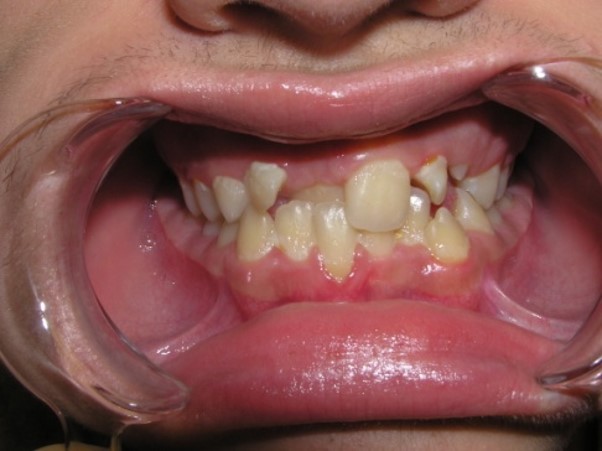
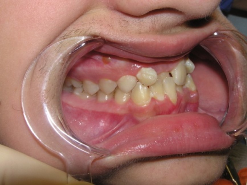
(one or both) - 61.35%. In 57% of children, there are erased areas on the
incisal and vestibular surfaces of palatal-displaced teeth as a result of the
functional action of antagonist teeth. With a slight overlap, abrasion of the
cutting edge and shortening of the abnormally located tooth are observed. In the
palatal location of the upper jaw incisors, flattening of the frontal area of
the upper jaw and lengthening of the frontal area of the lower jaw are
determined. In this case, exposure of the necks of the lower frontal teeth,
violation of their stability and inflammation of the gums may be observed, and
the clinic of catarrhal or atrophic gingivitis develops as a result of
functional trauma.
Etiology,
pathogenesis:
- heredity;
- harmful habits of the tongue
sucking, pencil, pen and other objects;
- disorder of respiratory and speech
functions, leading to underdevelopment of the jaws;
- mismatch of the size of the teeth
and jaw;
- crowding of the frontal
teeth;
- insufficient space in the dental
arch for the upper canines;
- delayed physiological change of
temporary teeth;
- trauma of the teeth, alveolar
process, jaws;
- macrodentia (absolute or
individual);
- presence of supernumerary
tooth;
- disorder of the teeth
clenching;
- micrognathia of the
jaw;
- mesial displacement of teeth
surrounding the abnormally located tooth;
- premature extraction of temporary
teeth without preventive measures;
- narrowing and shortening of the
dentition and apical base of varying degrees;
- underdevelopment of the upper jaw
frontal area;
- discrepancy between the width of
the dentition and apical base.
Functional disorders:
-
speech;
- biting
of food;
-
lengthening of chewing time;
-
disturbance of rhythm of chewing;
-
grinding type of chewing;
-
decrease of indicators of chewing efficiency;
-
disorder of lips closing;
-
abrasion of abnormally located teeth crowns;
-
diseases of periodontal tissues (localized, generalized);
-
temporomandibular joint disorder.
Aesthetic
changes:
- disorder of the lip
step;
- mismatch of the cosmetic center of
the face with the center of the upper and lower dental
arches;
- reduction of the upper
lip.
Forms of
anomaly:
- with space in the dentition;
- without space in the dentition;
- with concomitant anomalies of the dentition and bite;
- with compensated deficiency of
space in the dentition;
- with decompensated or complete
deficiency of space in the dentition.
All of forms can be combined with
different depths of overlap in the frontal area of the dental arches (slight,
medium and deep).
Sysoev N. P. (1975) proposed a
classification of the palatal position of the frontal teeth and identified 4
forms:
Form 1 – palatal position of the
frontal teeth when there is space in the dental arch of the upper jaw. Occurs in
42.4%
Form 2 – palatal position of the
frontal teeth with lack of space in the dental arch as result of the upper jaw
frontal area underdevelopment. Occurs in 29.6%
Form 3 – palatal position of the
frontal teeth, which is combined with protrusion of the lower jaw frontal teeth.
Occurs in 16.89% of cases.
Form 4 – palatal position of the
upper jaw frontal teeth with a lower teeth crowding. Occurs in 11.2%
Research methods:
1.
Clinical.
2.
Paraclinical.
X-ray:
-
orthopantomography;
-
computer tomography.
Biometric:
- dental
examination by methods:
Tonn
Bolton
Pont and
H. Linder, G. Hart
Korkhaus
Snagina
Little
Nance
Schwarz
Fuss
Schmuth
Facial
photometry.
Reopardodontography.
Principles of
treatment:
Preparatory period
-
psychotherapeutic preparation;
-
elimination of the risk factor (if it possible);
-
elimination of the etiological factor (if it possible);
-
sanitation of the oral cavity;
-
checking the state of oral hygiene, if necessary -
training;
-
prosthetic preparation (according to indications);
-
surgical preparation (according to indications).
Active
period of orthodontic treatment in temporary and mixed
dentition:
-
functional treatment methods (myogymnastics, massage,
etc.);
- use of
a spatula, wooden stick (if there is space);
-
appliances method of treatment taking into account the clinical situation of the
malocclusion (creation of space in the dental arch for an abnormally located
tooth: expansion of the dentition, lengthening of the dentition, distalization
of teeth, etc.);
-
leveling the depth of overlap (disconnection of dental arches) using a cap
fixation, occlusal overlays, an inclined plane;
-
normalization of functions (aesthetic, speech, chewing);
-
surgical treatment methods (serial sequential extraction of teeth by Hotz;
extraction of individual teeth; extraction of supernumerary tooth,
etc.);
-
stripping.
Active
period of orthodontic treatment in permanent
dentition:
- appliances treatment method taking into account the
clinical manifestation of the pathology (creation of space in the dental arc for
an abnormally located tooth: expansion of the dentition, lengthening of the
dentition, distalization of the teeth, etc.);
- normalization of functions
(aesthetic, speech, chewing);
- surgical treatment methods (extraction of individual teeth;
supernumerary tooth, compact osteotomy, insert of orthodontic implants,
etc.);
- stripping;
- prosthetic
treatment;
- functional treatment methods
(myogymnastics, massage, etc.);
- physiotherapeutic treatment
method: vacuum therapy, low-frequency therapeutic vibration, MRI-magnetic
resonance reflexotherapy, ultraphonophoresis, ultrasound, electrophoresis,
etc.
Retention
period of orthodontic treatment
- preservation of the achieved
result and prevention of relapse with the help of special
equipment;
- retainers (fixed,
removable);
- functional adaptation to the newly
created occlusion;
- extraction of the rudiments of third
molars (if necessary).