Distal teeth position
Distoocclusion of teeth (syn.:
dystoposition, distoanomaly, distal displacement of teeth) is the location of
teeth more distally in the dentition than they are normally located. An example
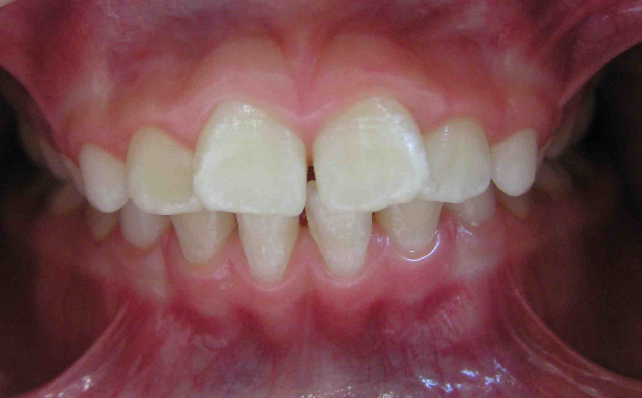
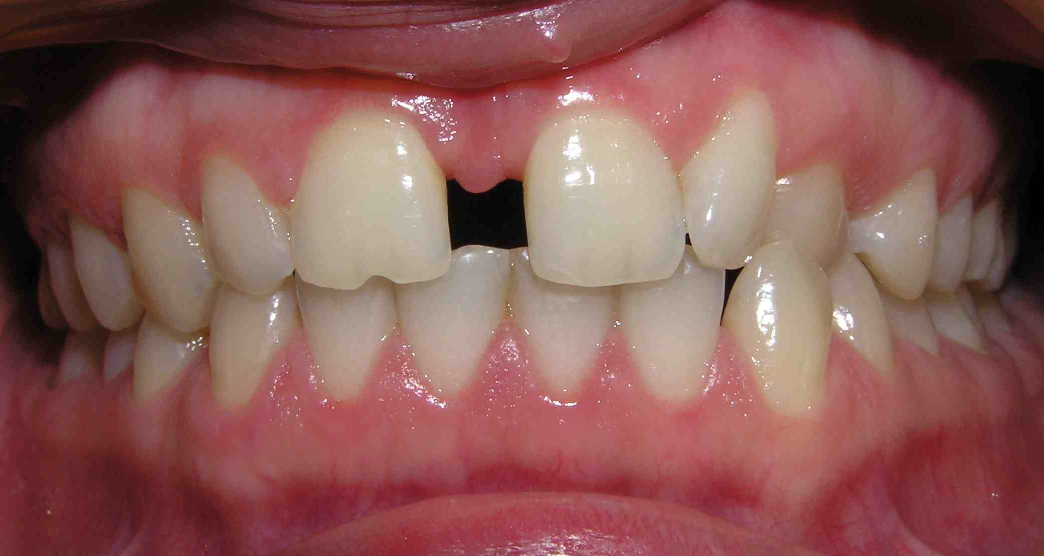
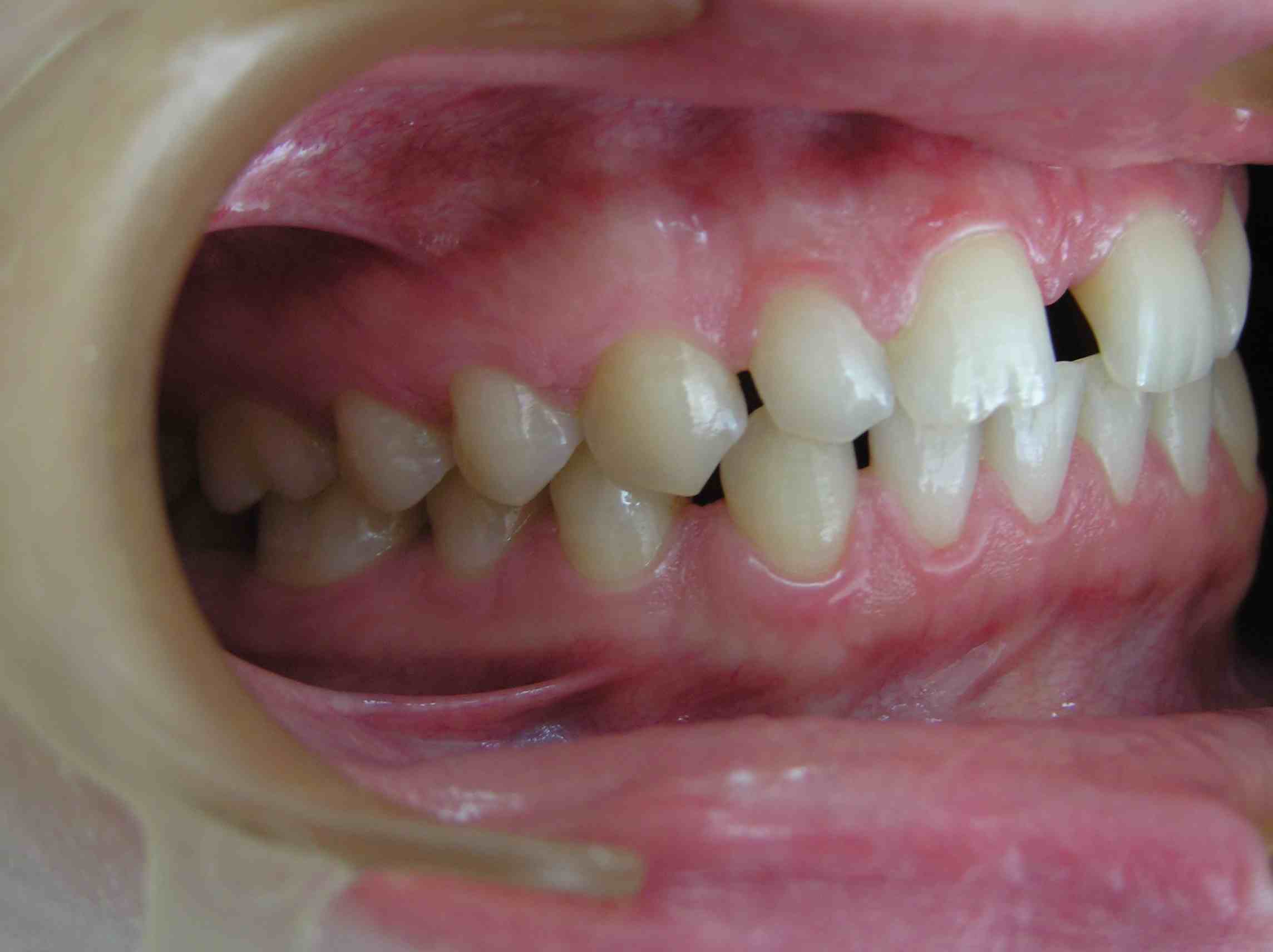
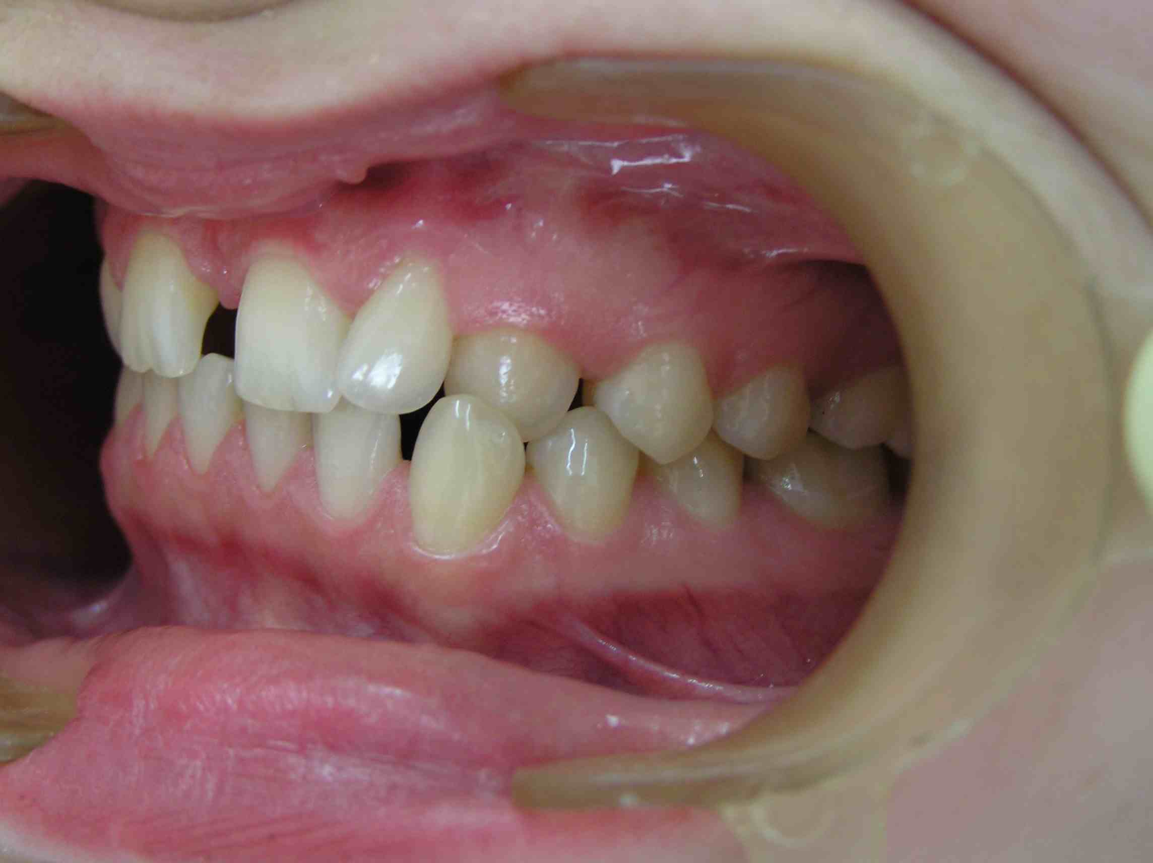
of the distal location of teeth is a diastema. Diastema in children during the
formation of the temporary bite, as a result of the low attachment of the
frenulum of the upper lip, is a common phenomenon. By the time the permanent
incisors erupt, the space between the temporary teeth increases, and after the
eruption of wider permanent teeth compared to the temporary teeth, the diastema
disappears. It should be noted that the presence of a diastema and a trema in
the III period of the temporary bite is considered as a sign of jaw growth, and
diastema and trema are physiological in nature and are not subject to
treatment.
Etiology,
pathogenesis:
- heredity;
- harmful habits of thumb sucking,
tongue, pencil, pen or other objects;
- disorders of swallowing, speech,
breathing, closing of lips;
- discrepancy of sizes of teeth and
jaw;
- trauma of teeth, alveolar process,
jaws;
- microdentia of lateral
incisors;
- mesiodent, supernumerary
tooth;
- adentia;
- retention of
tooth;
- disorder of tooth
formation;
- macrognathia of
jaw;
- premature extraction of temporary
teeth without preventive measures;
- expansion and lengthening of
dentition and apical base;
- discrepancy of width of dentition
and apical base.
Functional
disorders:
- speech;
- swallowing;
- disorder of closing of
lips;
- periodontal tissue diseases
(localized, generalized).
Aesthetic
changes:
- visualization of diastema when
talking and smiling.
Forms of
anomaly:
- symmetrical;
- asymmetrical
Research methods:
1.
Clinical.
2.
Paraclinical.
X-ray:
-
orthopantomography;
-
computer tomography.
Biometric:
- dental
examination by methods:
Tonn
Bolton
Pont and
H. Linder, G. Hart
Korkhaus
Snagina
Little
Nance
Schwarz
Fuss
Schmuth
Facial
photometry.
Reopardodontography.
Treatment principles:
Preparatory period
-
psychotherapeutic preparation;
-
elimination of the risk factor (if possible);
-
elimination of the etiological factor (if possible);
- oral
cavity sanitation;
-
checking the state of oral hygiene, if necessary -
training;
-
prosthetic preparation (according to indications);
-
surgical preparation (according to indications).
Active
period of orthodontic treatment in temporary and mixed
occlusion:
- functional treatment methods (myogymnastics,
massage, etc.);
- appliances treatment method taking
into account the clinical manifestation of the pathology (shortening of the
dentition, mesialization of the teeth);
- normalization of functions (aesthetic,
swallowing, speech, chewing);
- surgical treatment methods (plastic of lip
frenulum; extraction of supernumerary tooth, etc.).
Active
period of orthodontic treatment in permanent
occlusion:
- appliances treatment method taking
into account the clinical manifestation of the pathology (shortening of the
dentition, mesialization of the teeth);
- normalization of functions (aesthetic,
swallowing, speech, chewing).
- surgical treatment methods (extraction of
supernumerary tooth, compact osteotomy).
- prosthetic treatment;
- functional treatment methods (myogymnastics,
massage, etc.);
- physiotherapeutic treatment method: vacuum
therapy, low-frequency therapeutic vibration, MRI-magnetic resonance
reflexotherapy, ultraphonophoresis, ultrasound, electrophoresis,
etc.
Retention
period of orthodontic treatment
- preservation of the achieved result and
prevention of relapse using special equipment - retainers (fixed,
removable);
- functional adaptation to the newly created
occlusion;
- extraction of the rudiments of third molars
(if necessary).